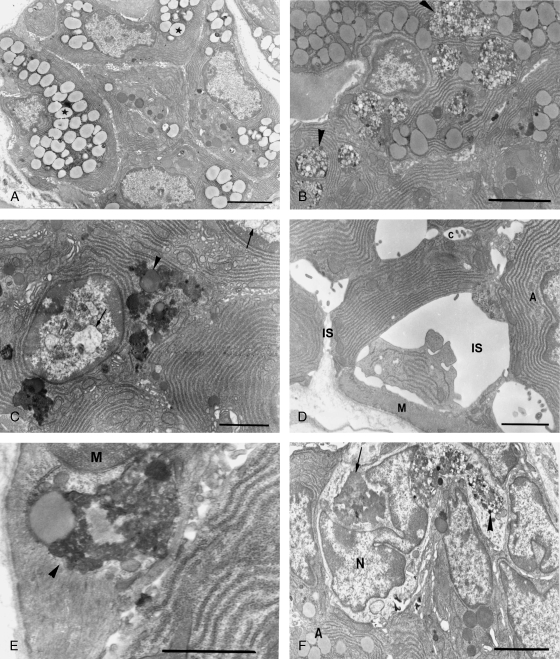
Fig. 3.
Ultrastructural changes of posterior deep lingual glands after hypoglossal denervation. (A) Three days after hypoglossal denervation, lipid droplets (asterisk) increased dramatically at the expense of secretory granules. Bar = 3.4 µm. (B) Seven days following axotomy, large bodies containing dense materials and numerous vesicles (arrowheads) are prominent. Bar = 4.0 µm. (C) Lipofuscin granules (arrowhead) are frequently found. Also note chromatin condensation and nuclear vacuolation (arrows). Bar = 2.5 µm. (D) The intercellular spaces (IS) around some acinar cells are dramatically enlarged. A: acinar cell; C: intercellular canaliculi; M: myoepithelial cell. Bar = 2.5 µm. (E) Numerous lipofuscin granules (arrowhead) are scattered in myoepithelial cells. M: myoepithelial cell nucleus. Bar = 2.0 µm. (F) Active phagocytes invading the acini. Note the lipofuscin granules (arrow) and large bodies containing dense materials and numerous vesicles (arrowhead) in the cell. A: acinar cell; N: nucleus of phagocyte. Bar = 6.5 µm.

