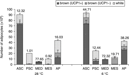
Fig. 3.
Density of TH-immunoreactive parenchymal nerve fibers (no. of fibers per 100 adipocytes) in the adipose organ of adult Sv129 female mice kept at 28 °C or 6 °C for 10 days. Numbers above columns are the average TH-positive fiber density in each depot (see also Table 1). ASC, anterior subcutaneous; PSC, posterior subcutaneous; MED, mediastinal; MES, mesenteric; AP, abdomino-pelvic. Mean ± SE. The bars represent SEM.

