Fig. 3.
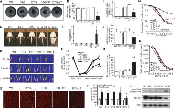
Impaired vascular function in mice with chronic Akt activation. (A) Sprouting of microvessels in Matrigel (arrows) from aortas derived from WT, single transgenic (STG), and double transgenic (DTG) mice with short-term (ST) and long-term (LT) Akt activation (n = 6 mice in each group). (B) Quantification of the microvessel sproutings from aortic ring. (C and F) Clinical score and photograph of leg gangrene in WT, STG, and DTG mice with and without short-term Akt activation and long-term Akt activation. Mice in the first four groups are normal, with a clinical score of 0. The DTG mice with long-term activation of Akt exhibited leg gangrene, with a clinical score of 2.9 (n = 6 to 8 mice in each group). (D and G) Laser Doppler images of hindlimb blood flow and quantification of blood flow in the lower limb after hindlimb ischemia in WT and mutant Akt mice (n = 6 to 8 mice in each group). (E and H) Representative images of microcapillaries in the hindlimb adductor muscle with quantification using Alexa 568–linked Isolectin B4 antibody (n = 6 to 8 mice in each group). (I) Measurements of eNOS activity in aortic lysates of WT and mutant Akt mice. (J) Nitrite accumulation from aortic endothelial cells (MAEC) derived from WT and mutant Akt mice. (K) Production of superoxide anion from mouse aorta as determined by lucigenin chemiluminescence assay. (L) Immunoblot analyses nitrotyrosinylation and expression of MnSOD in WT and mutant Akt mice. For nitrotyrosinylation of MnSOD, MnSOD was immunoprecipitated (IP) followed by immunoblotting with nitrotyrosine antibody. Results are presented as mean ± SD, n = 6 mice in each group. *P < 0.05 and **P < 0.01 when compared to other four groups. Relaxation of aorta to (M) acetylcholine (Ach) (endothelium-dependent) and (N) to sodum nitroprusside (SNP) in the presence of L-NAME (endothelium-independent) in WT, STG, and DTG mice, with and without short-term (ST) or long-term (LT) activation of Akt, and DTG mice with long-term Akt activation with rapamycin (DTG-LT/rapamycin). Relaxation is shown as percent of residual tone. (n > 6 aortas in each group, *P < 0.05 when compared to other groups, **P < 0.05 when compared to DTG-LT/rapamycin group.)

