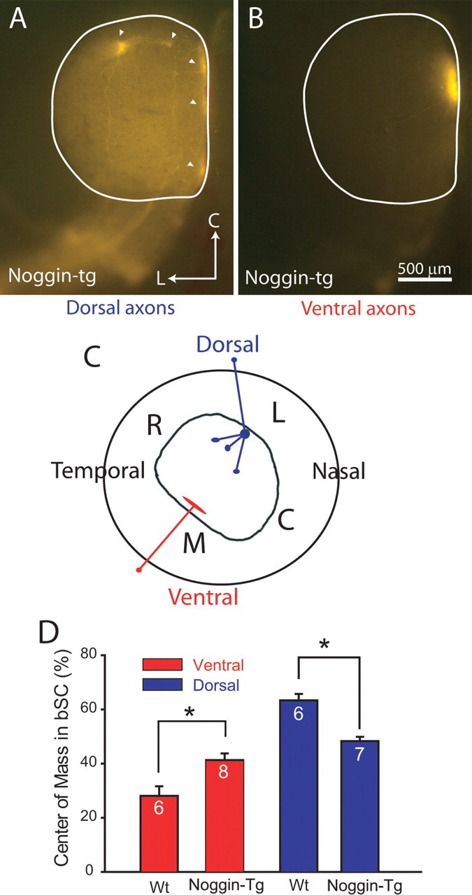
Figure 3.

Dorsal retinal axons in Noggin-transgenic mice are misplaced in the bSC at P8. A, Example of a dorsal retinal injection in a Noggin transgenic leading to multiple ectopic spots (arrowheads) at inappropriate locations in the SC. L, Lateral; C, caudal. B, Ventral retinal injections nearly always terminate normally in the SC. C, Summary diagram showing that dorsal injections lead to misprojections in Noggin transgenics, but ventral retinal injections are relatively normal. R, Rostral; L, lateral; M, medial; C, caudal. D, Summary quantification showing that the distribution of RGC axons in the bSC in Noggin transgenics is completely disturbed for dorsal injections (blue) and partially disturbed for ventral injections (red), so that dorsal (*p < 0.0001) and ventral (*p < 0.01) axons in Noggin transgenics are less confined to the lateral and medial sides of the bSC, respectively, than in WT mice. Error bars indicate SEM.
