Figure 9.

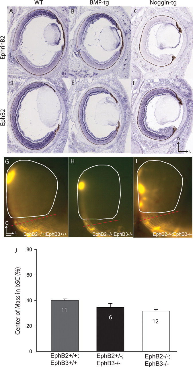
In situ hybridization reveals altered expression patterns of EphB2 and ephrinB2 in the retina of BMP-tg and Noggin-tg mice, but ventral axons are not missorted in EphB2/B3 mutant mice. A, In WT mice, ephrinB2 is expressed in a high-dorsal to low-ventral gradient. B, The ephrinB2 expression pattern is not altered in BMP-tg mice. C, EphrinB2 expression is dramatically suppressed in Noggin-tg mice. D, In WT mice, EphB2 shows a high-ventral to low-dorsal expression pattern. E, EphB2 expression is suppressed in BMP-tg, and no obvious gradient is present. F, EphB2 expression lacks a high-ventral to low-dorsal pattern in Noggin-tg mice, although average expression levels are normal. D, Dorsal; L, lateral. G–I, At P8, relative to controls (G), ventral RGC axons in EphB2+/−;B3−/− (H) and EphB2−/−;B3−/− mice (I) are not missorted in the brachium of the SC (G, H, red bar). C, Caudal. J, Summary quantification of center of mass for ventral injections in the bSC of WT and EphB2/B3 mutants. The sorting of EphB2/B3 double mutants is slightly better (with a smaller center of mass) than controls. Error bars indicate SEM. Scale bar: (in G) G–I, 100 μm.
