
Figure 1.
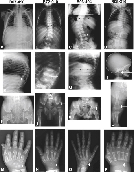
Radiographs of Individuals with SMDK with TRPV4 Mutations
Reference numbers are indicated above each column of radiographs. The ages of the subjects were 5 years (R07-490), 3 years (R72-010), 9 years (R03-404), and 8 months (R08-216).
(A–D) Anteroposterior (A/P) radiographs of the chest in four subjects, demonstrating the variability of the scoliosis from mild to more severe. The arrow in (C) shows the overfaced pedicles (lamina are seen medial to the vertebral edge).
(E–G) Lateral radiographs of the vertebrae showing flattened vertebrae (platyspondyly) and irregular margins.
(H) Lateral radiograph of the cervical vertebrae showing a small, irregular C1 vertebra (odontoid hypoplasia, arrow), a finding similar to metatropic dysplasia.
(I–K) A/P radiographs of the pelvis showing slightly flared ilia, wide epiphyseal plates, and mildly irregular acetabulae (arrows).
(L) A/P radiograph of lower extremity showing slightly widened metaphyses with normal epiphyses, somewhat reminiscent of metatropic dysplasia.
(M–P) A/P hand radiographs demonstrating the marked carpal ossification delay (arrows) for each subject's respective age.