
Figure 2.
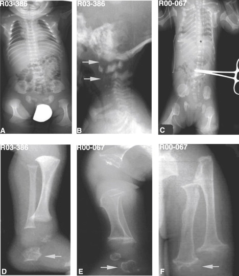
Radiographs of Metatropic Dysplasia in the Newborn Period
Reference numbers for the cases are indicated on each image.
(A and C) Body A/P radiographs in two cases showing severe platyspondyly (wafer-like), anterior and posterior rib cupping, widened ileum, and markedly flared proximal and distal femoral metaphyses (dumbbell-shaped femora).
(B) Lateral cervical spine radiograph demonstrating odontoid hypoplasia and hypoplasia of the cervical vertebral bodies with cervical kyphosis (arrows).
(D and E) Lateral lower-extremity radiograph showing characteristic irregular tali and calcanei (arrows) frequently seen in metatropic dysplasia.
(F) A/P radiograph of the upper extremity showing highly irregular metaphyses with popcorn appearance (arrow).