
Figure 3.
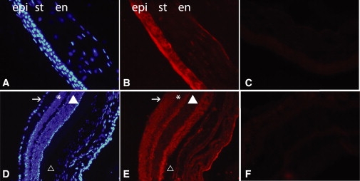
CNNM4 Immunostaining of Mouse Cornea and Retina
CNNM4 immunostaining of cornea (A–C) and retina (D–F) of 2-month-old mouse retina. In the cornea (B), CNNM4 is mainly localized in the epithelium (epi) surrounding the nuclei, in the keratocyte present in the stroma (st), and in the endothelium (en). (A) shows DAPI staining of the same slide, and (C) shows negative control without primary antibody. In the retina (E), immunostaining is mainly localized in the ganglion cell layer (indicated by the arrow), the inner and outer plexiform layers, and the outer segments of the photoreceptors. (D) shows DAPI staining of the same slide, and (F) shows negative control of without primary antibody. Symbols are used as follows: arrow, ganglion cell layer; asterisk, inner cell layer; filled triangle, outer cell layer; triangle, retinal pigment epithelium. The rabbit polyclonal antibody raised against amino acids 21–200 of human CNNM4 was used in a 1:200 dilution on 12 μm cryosections of eyes of 2-month-old C57Bl/6 mice. Secondary antibodies conjugated to Alexa Fluor 594 (Molecular Probes, Invitrogen) were diluted at 1:1000. Sections were stained with DAPI for visualizing nuclei before mounting in Cityfluor (Cityfluor Ltd.).