
Figure 2.
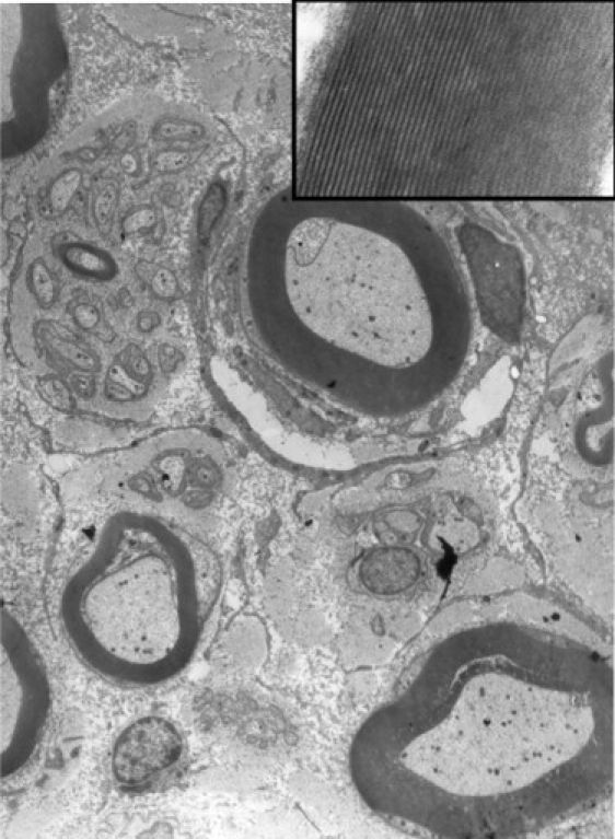
Electron Micrograph of the Sural Nerve Showing Normal Structure and Distribution of Myelinated and Unmyelinated Nerve Fibers
The inset shows details of a myelin sheath to show normal compaction and periodicity of myelin lamellae.
Official websites use .gov
A
.gov website belongs to an official
government organization in the United States.
Secure .gov websites use HTTPS
A lock (
) or https:// means you've safely
connected to the .gov website. Share sensitive
information only on official, secure websites.
Electron Micrograph of the Sural Nerve Showing Normal Structure and Distribution of Myelinated and Unmyelinated Nerve Fibers
The inset shows details of a myelin sheath to show normal compaction and periodicity of myelin lamellae.