
Figure 3.
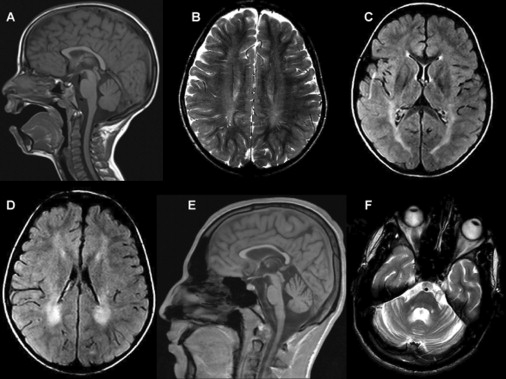
Brain MRI of Patient 2762 at 7 Years of Age and Patient 1211 at 18 Years of Age
(A) Sagittal T1 weighted image.
(B) Axial T2 weighted image.
(C and D) Axial FLAIR images showing prolonged relaxation times in the periventricular white matter, more prominent posteriorly, and to a lesser extent in the posterior limbs of the internal capsules. There is no significant loss of volume. Images in (A)–(D) are of patient 2762 at 7 years of age.
(E and F) Sagittal T1 weighted (E) and axial T2 weighted (F) images showing loss of volume of the corpus callosum and to a lesser extent of the cerebellum and pons. Images are of patient 1211 at 18 years of age.