
Figure 2.
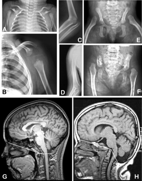
Radiographic Findings in Cousin Syndrome
(A, C, and E) Patient 1. (B, D and F) Patient 2. Skeletal features seen in the two patients include aplasia of the blade of the scapula, humeroradial synostosis, marked hypoplasia of the iliac bones, and dislocation of the femoral heads. (G) A sagittal section of a cranial MRI of a normal woman aged 19 years. (H) A corresponding section from patient 2 at age 12 years. From the tip of the odontoid process in the center of each panel, the arrows extend anteriorly to the frontal bone and posteriorly to the posterior margin of the foramen magnum. Caudal displacement of the occipital bone is evident in the patient; note also the redundant skin fold over the posterior aspect of the neck. The cranial and skeletal features are remarkably similar to those seen in Tbx15-ablated mice.5