
Figure 3.
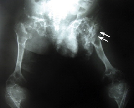
Antero-Posterior Radiography of the Pelvis, Femurs, and Knees of the 16-Year-Old Patient
Dense calcifications are projected over the left femoral head pubis and ischium (two arrows). The right acetabulum is flattened and irregular and shows a varus deformity of the femoral neck. The femurs and tibia metaphyses are widened.