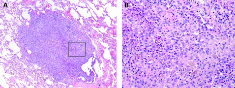
Figure 27.
Langerhans cell histiocytosis (LCH). LCH has variable morphology. The cellular form can suggest neoplasm (A). At higher magnification, the characteristic admixture of pale amphophilic Langerhans cells, lightly pigmented macrophages and eosinophils confirms the diagnosis (B). (A,B) H&E stain; (A) 40× original magnification; (B) 400× original magnification.

