
FIG. 8.
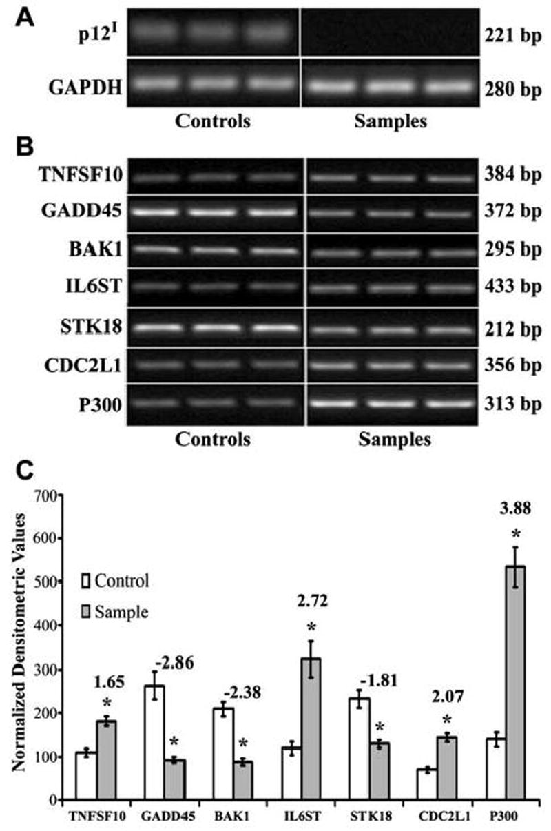
(A) RT-PCR demonstrating the expression of p12I-HA in primary CD4+ T cells 7 days postinfection with lentiviral vectors. Primary CD4+ T cells infected with sample vector express p12I whereas cells infected with control vector do not express p12I. RT-PCR for GAPDH was used as a control for the integrity of the message. (B) Semiquantitative RT-PCR demonstrating differential expression of selected genes in primary CD4+ T cells expressing p12I. Total cellular RNA was extracted 7 days postinfection with recombinant lentiviral vectors. Semiquantitative RT-PCR was performed on cDNA from 100 ng of total cellular RNA. RT-PCR was performed with triplicate samples and controls. GAPDH was used as a control for the integrity of the message. (C) Graph demonstrating densitometric analysis of semiquantitative RT-PCR of selected genes in primary CD4+ T cells expressing p12I. Fold difference between control and sample is given at the top of the column for each gene. Results are expressed as means with standard error (SE) from a minimum of triplicate experiments. BAK1, GADD45, and STK18 were downregulated whereas p300, CDC2L1, TNFSF10, and IL6ST were upregulated by p12I. Statistical analysis was performed using Student t test. *p < 0.05.