
Figure 1.
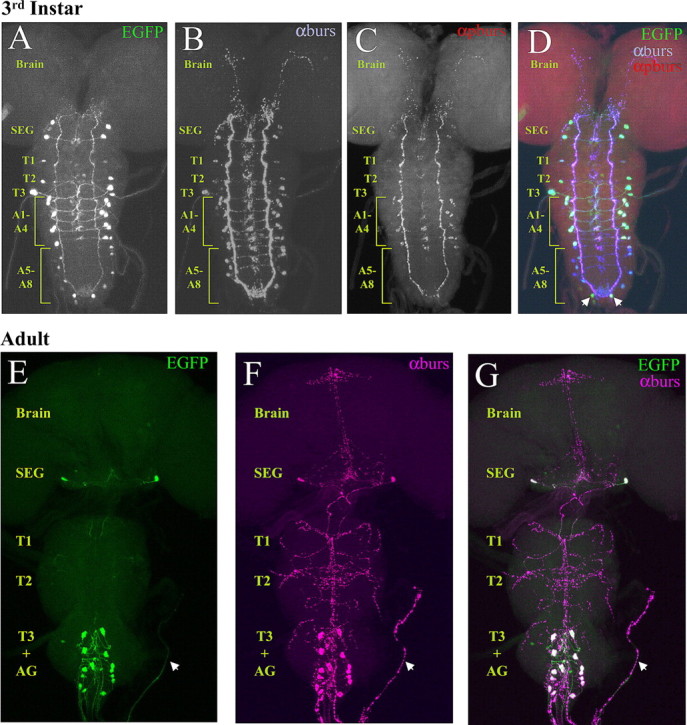
Expression of Burs-Gal4 mimics the expression pattern of bursicon α-subunit (burs) in both larval and adult nervous systems. Confocal images of nervous system whole mounts from Burs-Gal4>UAS-EGFP animals at the third larval instar and adult stages. A–D, Larval nervous system expressing EGFP under the control of Burs-Gal4 (A), triple labeled with antibodies to both burs (B) and pburs (C). In the merged image (D), the green, blue, and red channels represent EGFP, burs, and pburs labeling, respectively. Neurons immunopositive for burs are consistently labeled with EGFP. These include the following: two prominent pairs of neurons in the subesophageal ganglion; one pair in each of the first two thoracic neuromeres and two pairs in the third; two pairs of neurons in each of the first four abdominal neuromeres; and one to two pairs in each of the remaining abdominal neuromeres. Arrows indicate two neurons in the terminal abdominal segment that are labeled by Burs-Gal4, but which do not express burs. In contrast to burs, pburs expression is prominent only in the first four abdominal segments, with weak labeling in some of the burs-immunopositive subesophageal neurons. SEG, Subesophageal ganglion; T1–3, thoracic neuromeres 1–3; A1–8, abdominal neuromeres 1–8. E–G, Adult nervous system from a newly eclosed Burs-Gal4>UAS-EGFP animal expressing EGFP (E, green), double labeled with burs antibodies (F, magenta). G, Merged image shows complete correspondence of labeling, with overlapping labels appearing as white. Whole mounts are oriented symmetrically along the rostral-caudal axis with the midline of the nervous system in the center. Arrow, An intact section of abdominal nerve. AG, Abdominal ganglion.