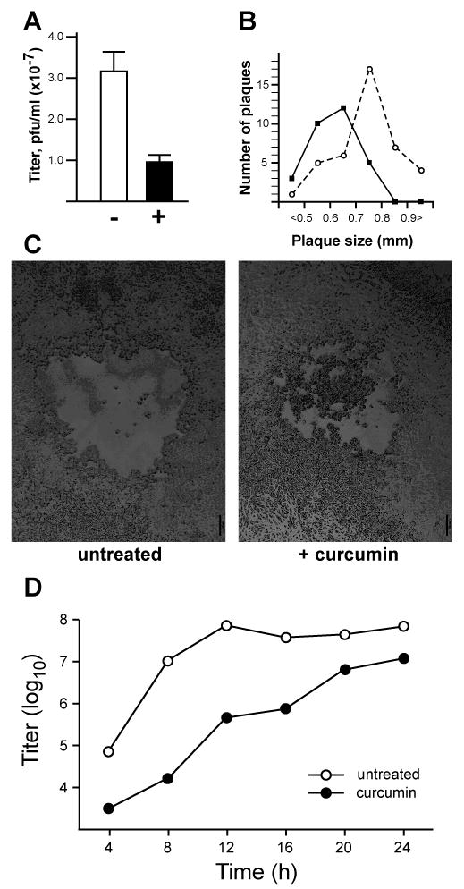
Figure 1. Effects of curcumin on HSV-1 plaque formation and viral replication.

(A) Plaque assays were performed in Vero cells pretreated (+) with 10 μM curcumin or untreated (-) for 2 hours and infected with serial dilutions of a stock preparation of HSV-1. 10 μM curcumin was also present in the viral inoculums and overlay agar media of treated samples. Error bars represent the standard deviation among triplicate assays at two viral dilutions. (B) Plaque sizes in the absence (open circles) or presence (closed circles) of curcumin were measured by averaging randomly chosen vertical and horizontal diameters from plaques. The numbers of plaques within various size ranges (from less than 0.5 mm to greater than 0.9 mm) are shown. (C) Representative plaques from plaque assays performed in the absence (left panel) and presence (right panel) of 10 μM curcumin. Scale bar: 100 μm. (D) Single-step growth curves were performed in Vero cells infected with HSV-1 at an MOI of 1 pfu/cell. Virus titers in samples collected at 4 h intervals were assayed in triplicate on Vero cells. Closed circles represent curcumin-treated samples; open circles represent parallel cultures of untreated cells.
