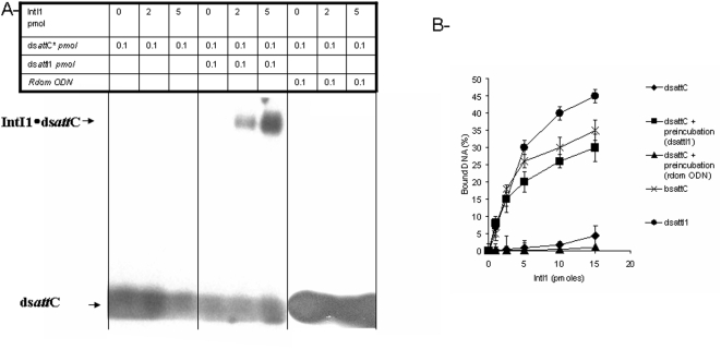
Figure 3. Cooperative binding of IntI1 to attI1/attC sites.
A- In vitro DNA binding of IntI1 with double-stranded attC fragment in presence or not of attI1 site. Free 5′ 32P radiolabeled dsDNA attC fragments containing recombination sites (0.1 pmoles) were incubated at 4°C for 20 min with purified IntI1 (0–5 pmoles) after or without preincubation with dsattI1 (0.1 pmole) or random ODN (0.1 pmole) at 4°C for 20 min. Products were then loaded on 1% agarose gel and electrophoresis was run at 50 V, for 2 hours at 4°C. B- Effect of preincubation with different att fragments on in vitro DNA binding of IntI1. Free 5′ 32P radiolabeled dsDNA fragments containing recombination sites dsattC, dsattI1, or bsattC (0.1 pmoles) were incubated at 4°C for 20 min with purified IntI1 (0–5 pmoles) after or without preincubation with dsattI1 (0.1 pmole) or with a random ODN (0.1 pmole) at 4°C for 20 min. DNA binding was measured by quantification of gel shifted bands using DNAJ software and also filter binging assay as described in materials and methods section. The percentage of bound DNA was then plotted in the graphic B. Results are the mean±standard deviation (error bars) of three independent experiments.

