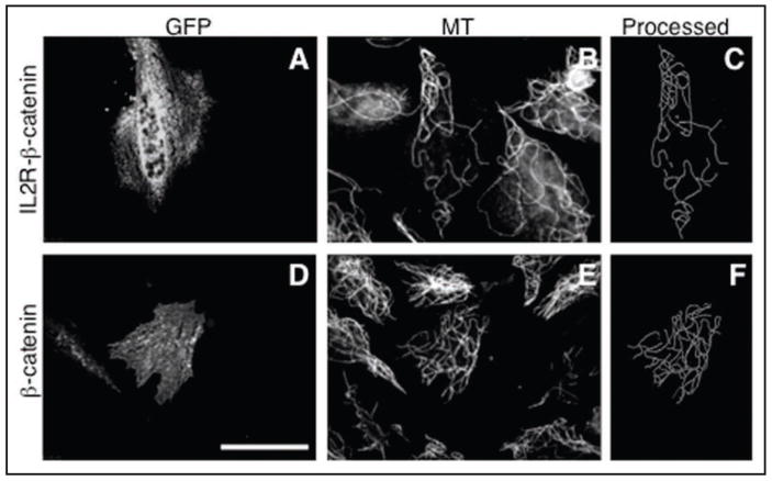
Figure 4.
Effect of expression of membrane-targeted and cytoplasmic β-catenin on the microtubule density in the centrosome-lacking cytoplasts. Upper: cells transfected with membrane targeted IL2R-β-catenin-GFP chimera (A–C), lower: cells transfected with the β-catenin-GFP (D–F). From left to right: GFP fluorescence in cytoplasts produced from transfected cells (left), microtubule staining of the same field of view (middle), processed microtubule image of the transfected cytoplasts (right). Scale bar represents 10 μm.

