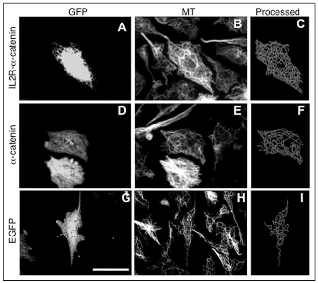
Figure 5.
Effect of membrane targeted α-catenin expression on microtubule density in the centrosome-lacking cytoplasts. Upper: cells transfected with membrane targeted IL2R-α-catenin-GFP chimera (A–C), middle: cells transfected with the α-catenin-GFP (D–F), lower: cells transfected with the enhanced GFP alone (G–I). From left to right: GFP fluorescence in cytoplasts produced from transfected cells (left), microtubule staining in the same field (middle), processed microtubule image of the transfected cytoplasts (right). Scale bar represents 10 μm.

