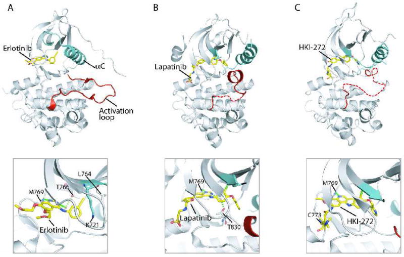
Figure 3.
Structures of EGFR kinase domain bound to small molecule kinase inhibitors. (A) Erlotinib - EGFR structure [15]. (B) Lapatinib - EGFR structure [22]. (A) HKI-272 - EGFR structure [72]. The upper panels show the entire EGFR kinase domain with inhibitor bound, revealing the active (A) and inactive (B and C) conformations of EGFR. The lower panels show close-up views of the bound inhibitors.

