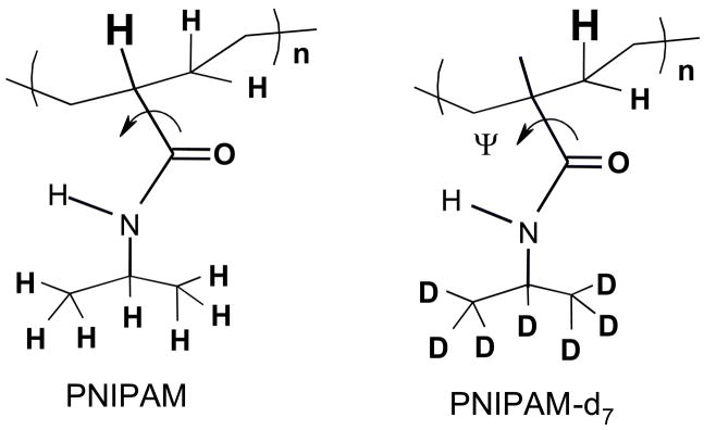
Figure 3.
The structure of PNIPAM and its deuterated isotopomers, d7-PNIPAM. The Cα-hydrogen that gives rise to the resonance enhanced CαHb band at ~1387 cm−1 is highlighted. The curved arrow highlights the Ψ dihedral angle rotation around the Cα-C(O) bond. The AmIII band frequency is sensitive to the Ramachandran Ψ angle rotation around the Cα-C(O) bond.

