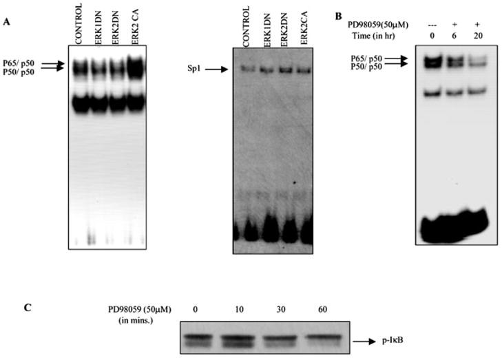
Fig. 7. ERK is involved in the NF-κB binding to DNA.
A, Hs294T melanoma cells were transfected with either empty vector or ERK dominant negative or dominant active constructs. Twenty hours after transfection, cells were placed in serum-free medium. Forty-eight hours after transfection, cells were harvested, and nuclear extracts were made. 10 μg of nuclear extracts were incubated with 32P-labeled NF-κB or Sp1 oligonucleotide probe in the presence of 1–2 μg of poly(dI-dC). The reaction mixture was electrophoresed on a 6% native gel, which was dried and processed for autoradiography. The arrow indicates the specific complexes associated with the NF-κB and the Sp1 probe. The EMSA shown is representative one of three different experiments. Results were quantitatively similar in all three experiments. B, Hs294T cells were incubated with PD98059 (50 μM) for the indicated time periods, nuclei were extracted, and an NF-κB EMSA was performed as described for A. C, Hs294T melanoma cells were preincubated with proteosome inhibitor MG115 and cycloheximide for 2 h and then treated with PD98059 (50 μM) for 10, 30, and 60 min, respectively. The cells were then lysed using radioimmune precipitation buffer, and protein was estimated. 50 μg of protein were loaded in each lane, and SDS-PAGE analysis under reducing conditions was performed. The effect of PD98059 on phosphorylation of IκB was determined using phosphospecific Iκβ antibody.

