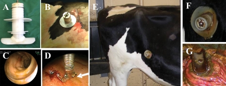
FIG. 1.
Pictures showing the modified cannula used in the study (A), appearance of cannula after surgery (B), endoscopic field showing no inflammation present at the time of inoculation with M. avium subsp. paratuberculosis (C), and collection of pinch biopsy (D). Other images show the status of cannula 8 months postsurgery just before necropsy. Inflammation at the site of implantation was kept at a minimum by keeping the site clean (E and F). Inspection of the internal portion of the cannula showed minimal inflammation (G).

