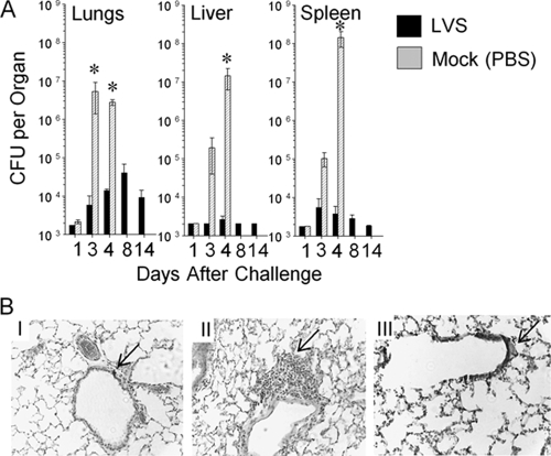
FIG. 7.
Oral LVS vaccination leads to a reduction in bacterial replication and an increase in inflammatory response following pulmonary SCHU S4 challenge. Groups of BALB/c mice (n = 5) were either vaccinated orally with 103 CFU of LVS or mock immunized with PBS. Three weeks later, mice were challenged i.n. with 130 CFU of F. tularensis SCHU S4. (A) Mice were sacrificed at different time points (1, 3, 4, 8, and 14 days) after challenge, and various organs were removed. Bacterial numbers were enumerated by homogenization of whole individual organs and serial dilution plating. *, differences in bacterial numbers between LVS- and mock-immunized mice were significant at a P value of <0.05 (statistical power, 0.999 with an alpha of 0.50). (B) Mice were sacrificed 3 days after challenge, and lungs were collected for hematoxylin-and-eosin analyses. (I) Mock-vaccinated, SCHU S4-challenged mice. The arrow indicates the absence of peribronchiolar cellular infiltration. (II) LVS-vaccinated, SCHU S4-challenged mice. The arrow indicates the presence of peribronchiolar mononuclear lymphocytic infiltration. (III) Mock-vaccinated mice, no challenge. The arrow indicates normal bronchiolar architecture. Images are shown at a magnification of ×50. Results are representative of two separate experiments.

