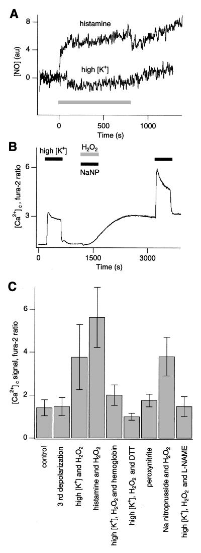
Figure 5.
NO is involved in potentiation of Ca2+ signaling. (A) NO level measured by using DAF-2 in PC12 cells during application of either 100 μM histamine or K+ depolarization (shown by bar). Representative results are from the series of four experiments for each treatment. (B) Ca2+ signal in PC12 cells in response to K+ depolarization before, during, and after simultaneous application of 100 μM NaNP and 0.03% H2O2. (C) Ca2+ signal amplitude in undifferentiated PC12 cells in response to K+ depolarization after the treatment listed. Control, K+ depolarization in naïve cells. Concentrations used were 35 mM K+, 0.03% H2O2, 100 μM histamine, 0.1 mM hemoglobin, 5 mM DTT, 100 μM peroxynitrite, 100 μM NaNP, and 12 mM l-NAME. For each experiment, n = 4–7.

