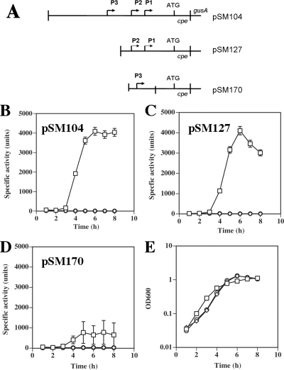
FIG. 9.
Expression of cpe-gusA fusions in C. perfringens wild-type and mutant strains. (A) Schematic diagram of the C. perfringens cpe translational fusions to E. coli gusA. (B to D) Expression from the cpe promoters in the indicated plasmids was measured by the specific activity of β-glucuronidase in samples collected during an 8-h time period after inoculation of wild-type SM101 (squares) and mutants KM1 (sigK mutant) (diamonds) and KM2 (sigE mutant) (circles) into sporulation medium. (E) Representative growth curves of strains SM101 (squares), KM1 (sigK mutant) (diamonds), and KM2 (sigE mutant) (circles) containing pSM104 in sporulation medium (growth patterns with strains containing the other plasmids were very similar).

