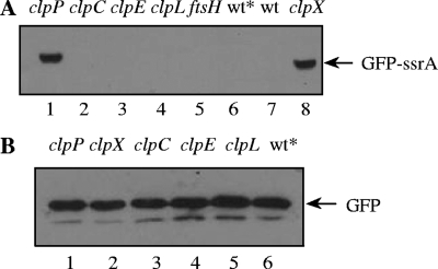
FIG. 2.
Stabilization of GFP-SsrA in clpX and clpP mutants. (A) Strains expressing gfp-ssrA in clpP (CP1904), clpC (CP1906), clpE (CP1907), clpL (CP1908), ftsH (CP1910), or clpX (CP1905) mutant backgrounds were harvested, lysed, and loaded in lanes 1 to 5 and 8 of a sodium dodecyl sulfate-polyacrylamide gel electrophoresis gel. CP1903, expressing gfp-ssrA in the wild-type background (wt*), and the wild-type with no gfp insertion at the aga locus (wt) were loaded in lanes 6 and 7, respectively. (B) Strains expressing untagged gfp in clpP (CP1912), clpX (CP1913), clpC (CP1914), clpE (CP1915), or clpL (CP1916) mutant backgrounds or in the wild-type background (wt*; CP1911) were harvested, lysed, and loaded in lanes 1 to 6. Strains expressing gfp-ssrA or untagged gfp in protease-proficient backgrounds are represented as wt*. Cultures were grown to an optical density at 550 nm of 0.25 in a casein hydrolysate yeast extract medium (13) supplemented with raffinose (1 g/liter) for maximal induction of the aga locus. Cell pellets were resuspended and heated in a 1/100 volume of lysis buffer (100 mM Tris-HCl [pH 6.8], 4% sodium dodecyl sulfate, 0.2% bromophenol blue, 20% glycerol, and 200 mM dithiothreitol) for determination of GFP or GFP-SsrA by Western blotting with anti-GFP antibody (Roche). After sodium dodecyl sulfate-polyacrylamide gel electrophoresis and transfer to polyvinylidene difluoride membranes, the membranes were probed with a mouse anti-GFP primary antibody (1:1,000; Roche) and with an anti-mouse immunoglobulin G secondary antibody conjugated to horseradish peroxidase (1:10,000; GE). Detection was performed with an enhanced chemiluminescence substrate (ECL Plus; GE) and Hyblot CL film (Denville Scientific) with exposures between 10 and 600 s.

