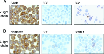
FIG. 1.
PEL cells express low levels of cIg. Cytospins of PEL cell lines were analyzed for expression of κ (A) and λ (B) light chains by immunohistochemistry. BC1 showed very faint κ expression. BC3 was negative for κ light chains but showed rare λ-positive cells. A few BCBL1 cells were positive for λ light chains. BJAB and Namalwa cells were used as positive controls for κ and λ light chain expression, respectively. Experiments were performed at least three independent times.

