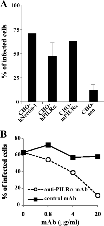
FIG. 5.
PRV infection of CHO cells expressing PILRα. (A) CHO-hNectin-1, CHO-hPILRα, CHO-mPILRα, and CHO-neo cells were infected with PRV at an MOI of 5. At 8 h postinfection, infected cells were fixed, permeabilized, stained with anti-EP0 antibody, and analyzed by flow cytometry to determine the percentages of infected cells. The mean and standard deviation from three independent experiments is shown for each cell type. (B) CHO-hPILRα cells were infected with PRV at an MOI of 5 in the presence of various concentrations of anti-PILRα MAb or control MAb. At 8 h postinfection, infected cells were stained with anti-EP0 antibody and analyzed by flow cytometry to determine the percentages of infected cells.

