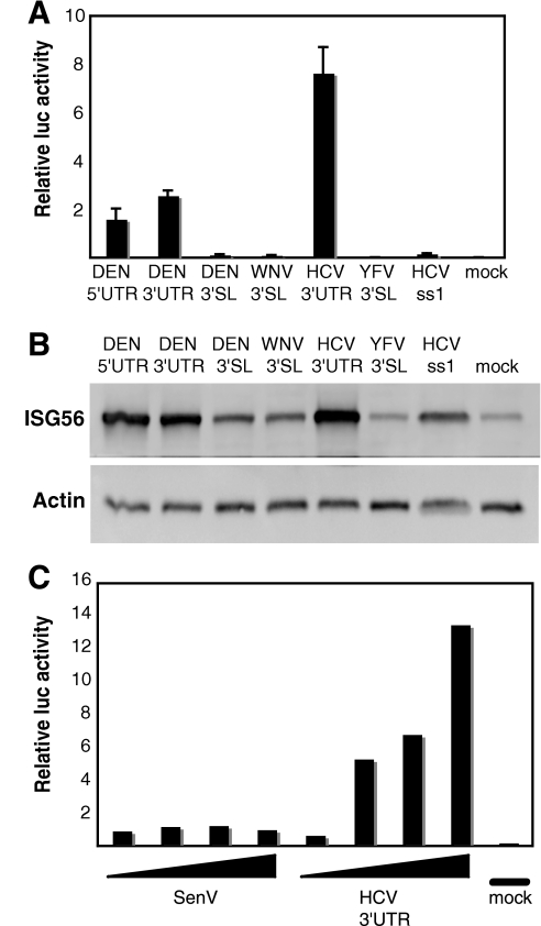
FIG. 1.
IFN-β induction potentials of HCV and flavivirus UTR RNAs. (A) Twenty-four hours after being plated, Huh7 cells were cotransfected with plasmids encoding firefly or Renilla luciferase under the control of the IFN-β promoter or constitutive cytomegalovirus promoter, respectively. Following a 24-hour further incubation period, the cells were mock transfected or transfected in triplicate with equal numbers of moles of renatured in vitro-transcribed viral 5′- or 3′-UTR or 3′-SL RNAs. Twenty-four hours later, the cells were lysed, and aliquots of the extracts were analyzed using a dual-luciferase assay. The firefly luciferase light unit values were divided by the Renilla light units (transfection efficiency control) to generate the relative luciferase (luc) value. The bars show average relative luciferase values plus standard deviations. (B) Twenty-eight hours after viral-RNA transfection, the cells were lysed and analyzed by SDS-polyacrylamide gel electrophoresis and immunoblotting for ISG56 and actin. (C) Huh7 cells were transfected or infected with increasing amounts of HCV 3′-UTR RNA (50 ng, 250 ng, 650 ng, and 1 μg) or Sendai virus (SenV) (50, 100, 250, or 500 hemagglutinin units), and IFN-β reporter activation was measured 24 h later as described in the legend to Fig. 1A.

