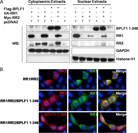
FIG. 4.
EBV RR1, RR2, and BPLF1 1-246 localize in the cytoplasm of 293T cells. (A) 293T cells were transfected with EBV RR1, RR2, and BPLF1 1-246 for 48 h. Fractionated extracts were analyzed by Western blotting (WB). The notation above the blots indicates the transfecting plasmids. The notation to the right indicates the protein blotted against. GAPDH (glyceraldehyde-3-phosphate dehydrogenase) served as a cytoplasmic marker, and histone H1 served as the nuclear marker. (B) 293T cells were transfected with HA-tagged RR1, c-myc-tagged RR2, and FLAG-tagged BPLF1 1-246 as indicated to the left. Immunofluorescence staining was performed with antibody against HA, myc, and FLAG. Nuclear DAPI staining is shown in blue. All panels represent ×100 magnification. Top panels depict cells transfected with RR1 and RR2 stained for RR1 and RR2. Middle and bottom panels depict cells transfected with RR1, RR2, and BPLF1 1-246 stained for RR1 and RR2 or RR1 and BPLF1 1-246, respectively. Merged images are shown to the right.

