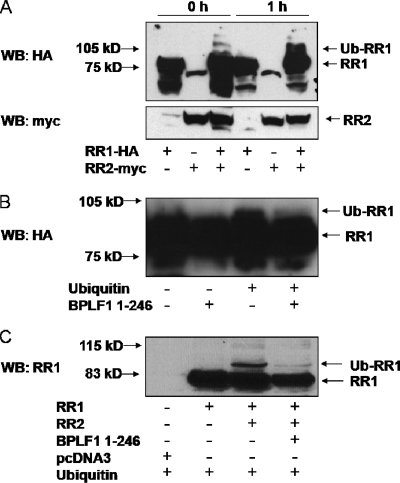
FIG. 5.
EBV RR1 is ubiquitinated in vitro and in vivo and is deubiquitinated by BPLF1 1-246. (A) 293EBV+ cells were transfected with RR1, RR2, or both as indicated. Cells were harvested after 48 h, and lysates were mixed with reaction buffer containing rabbit reticulocytes and exogenous Ub. Samples were then incubated at 37°C for the indicated amounts of time and subjected to Western blotting (WB). The top panel shows probing with HA antibody, and the bottom panel shows probing with c-myc antibody. The locations of RR1, RR2, and ubiquitinated RR1 (Ub-RR1) bands are indicated by arrows. (B) 293EBV+ cells were transfected with EBV RR1 and RR2. Cells were harvested after 48 h, lysates were mixed with reaction buffer, and exogenous Ub was added where indicated, followed by incubation of samples at 37°C for 1 h. Purified BPLF1 1-246 was added to the indicated samples, and the reaction was continued for another hour at 37°C. Arrows note the location of RR1 and ubiquitinated RR1. The blot was probed with anti-EBV EaR (RR1) and is overexposed to show RR1 ubiquitination. (C) 293T cells were transfected with RR1, RR2, and BPLF1 1-246 as indicated. All samples were additionally transfected with HA-tagged Ub and subjected to Western blotting against RR1. Arrows note the location of RR1 and ubiquitinated RR1.

