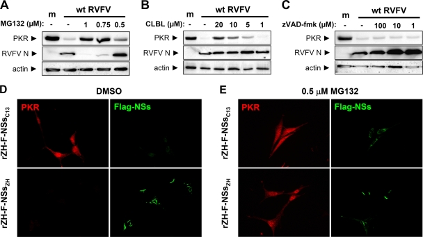
FIG. 6.
Involvement of the proteasomal pathway. (A to C) Western blot analysis. IFNAR−/− MEFs were seeded in six-well dishes and left uninfected (m) or infected with wt RVFV at an MOI of 5. After 1 h of infection, fresh medium with the indicated concentrations of MG132 (A), CLBL (B), or the pan-caspase inhibitor zVAD-fmk (C) was added directly onto the cells. As a control, the medium in untreated cells was supplemented with equivalent amounts of dimethyl sulfoxide (DMSO). After 12 h of incubation, cell lysates were prepared and subjected to Western blot analysis as described for Fig. 4. (D and E) Immunofluorescence analysis. IFNAR−/− MEFs grown on coverslips were infected (MOI of 5) with recombinant RVF viruses expressing the NSs proteins of either Clone 13 or the wt strain ZH548, each fused to a C-terminal Flag tag. During the incubation period of 16 h, cells were either treated with DMSO (D) or with 0.5 μM MG132 dissolved in DMSO (E). Then, cells were fixed and analyzed by indirect immunofluorescence with antibodies directed against PKR (red) or the Flag portion of the fusion proteins (green).

