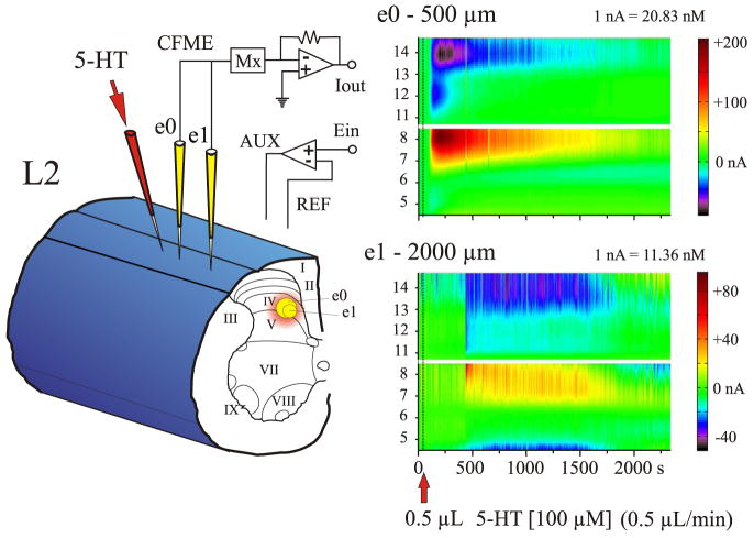
FIG. 2.
Experimental trial of 5-HT microinjection into the spinal cord. Left: 2 CFMEs, e0 and e1 (yellow), and the 5-HT micropipette (red) were lowered into lamina IV of the L2 spinal segment. e0 was 500 μm and e1 was 2,000 μm away from the 5-HT micropipette. Right: color representations showing serotonergic redox signals at e0 and e1 after microinjection of 0.5 μl (dashed line) of 100 μM 5-HT over 1 min. Note that 5-HT was detected sooner and at a much higher concentration by e0 compared with e1.

