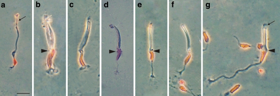
Fig. 2.
Photomicrographs showing isolated adult photoreceptors from P1–P15 chick retinas with their nucleus stained by the Feulgen method (red or purple). a–c, e–g Phase contrast. d Transmitted light with the condenser diaphragm nearly closed to discern the cell contour. a Rod. b Double cone (arrowhead position of OLM) with the principal member (left) and the accessory member (right). c Principal cone similar to that in Fig. 1k. Note that its nucleus extends from the OLM to the synaptic pedicle. d Accessory cone showing a portion of its nucleus outside the OLM (arrowhead). e, f Straight cones with the nucleus in the outer and middle positions, respectively (arrowhead position of OLM). g Oblique cone (arrowhead position of OLM). Arrow points to outer segment. Bar 10 μm

