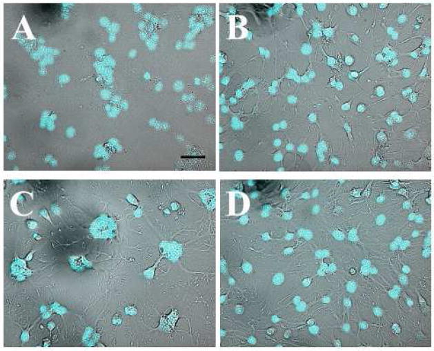
Figure 6.
Disturbance of actin structures with jasplakinolide, latrunculin, and cytochalasin D does not block syncytium formation.
Latrunculin A (panel B), jasplakinolide (panel C), and cytochalasin D (panel D) were used respectively at final concentrations of 2 μM, 3 μM, and 5 μM with a preincubation time of 30 min. After low pH application, the cells were kept in the presence of the reagents until scoring syncytia. Panel A shows the positive control, where the syncytium totally covered the field observed. B–D. Syncytia are still formed in the presence of actin-modifying agents. Scale bar, 50μm.

