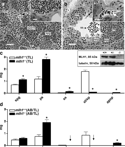
Fig. 1.
Testis histology of mlh1−/− zebrafish. a, b Cross section of seminiferous tubule in mlh1+/+ and mlh1−/− zebrafish, respectively, showing spermatogenic cysts with various types of germ cells: different types of spermatogonia (spg), primary spermatocytes (ps), secondary spermatocytes (ss), spermatids (st), spermatozoa (sperm), and apoptotic cells (A). Note the abnormal spermatogenesis in the mutant with many secondary spermatocytes having been released into the lumen, and very few sperm being produced (b, inset, arrow), whereas numerous spermatozoa are visible in the tubular lumen and efferent ducts of wild-type males (a, inset). Bars 25 μm (a, b), 10 μm (insets) c Morphometric analysis of testis sections from fish with TL background. The mutant shows significantly higher amounts of spermatogonia (spg), primary spermatocytes (ps), secondary spermatocytes (ss), and apoptotic cells (apop) than the wild-type. On the other hand, spermatids and spermatozoa (st/sp) are significantly reduced. Inset: Western blot of testes proteins stained with antibodies for MLH1 (85 kDa) and tubulin (50 kDa). MLH1 protein is completely absent in mlh1−/− testes (−/−) but detectable in wild-type (+/+) and heterozygous (+/−) males. d Morphometric analysis of testis sections from wild-type and mlh1−/− mutant zebrafish on an AB/TL background (n = 6 for both genotypes). The mutant shows an increased mass of spermatogonia (not significant), primary spermatocytes, and apoptotic cells. Secondary spermatocytes, spermatids, and spermatozoa were completely absent (arrows). Significant differences between wild-type and mutant genotypes are indicated (*P < 0.05)

