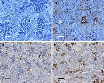
Fig. 2.
Testis immunohistochemistry of mlh−/− zebrafish. a, b TUNEL staining of mlh1+/+ and mlh1−/− fish, respectively. Large numbers of apoptotic cells can be seen in mutant testis (brown). Wild-type testis exhibits a low incidence of apoptosis. c, d Histone H3 staining of cells in metaphase from mlh1+/+ and mlh1−/− fish, respectively. Note the higher incidence of stained cells in mutant testis indicating a higher number of mitotic and meiotic cells in metaphase. Bars 25 μm (a, b), 50 μm (c, d)

