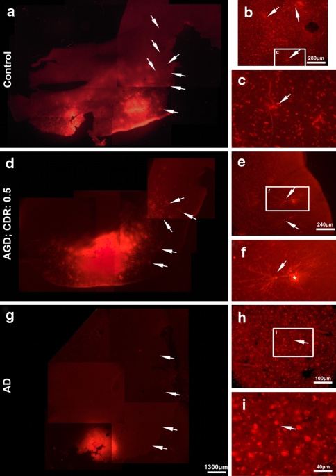
Fig. 2.
Disconnection of the entorhinal–temporal association fiber tract in an AD case but not in cognitively impaired AGD and control cases. a DiI-tracing of entorhinal–temporal connections in the control case (case No. 2) exhibited DiI-labeled structures in area 35 of the temporal neocortex (arrows). b Detailed view into the temporal cortex of area 35 of the control case. Here, a group of three neurons (arrows) was labeled. One neuron (boxed area) is enlarged in c. The cell soma and the basal dendrites were traced (arrow). d A similar pattern of traced neurons was seen in the cognitively impaired AGD case (arrows; case No. 3). e The detailed view into area 35 showed a cluster of two neurons (arrows). One neuron (boxed area) is enlarged in f showing the soma with the dendritic tree (arrow). Artificially stained material was in contact with the neuron and is indicated by an asterisk. g No DiI-labeling of association fibers in the temporal cortex of the AD case (arrows; case No. 6). h Only single neurites (arrow in h and i) were detectable in the entorhinal cortex (boxed area) and are enlarged in i and Fig. 3c. Calibration barg valid for a, d, and g; and h valid for c, f, and h

