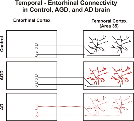
Fig. 5.
Schematic representation of temporal–entorhinal connectivity in control, AGD and AD brain. In aged controls with minimal AD-related changes temporal and entorhinal cortices are connected. Typical pyramidal neurons project into the entorhinal cortex. In AGD temporal and entorhinal cortices are connected but the projecting pyramidal neurons display dendritic alterations (marked in red). Axonal disconnection is seen in AD. Neurons, which once were connected, are no longer detectable with DiI-tracing (scattered neurons marked in red). The red-labeled parts of the association neurons were obviously altered in AGD and AD, respectively. Disconnection in AD can most likely be explained as the result of neuronal loss [5, 13, 15, 26, 33]

