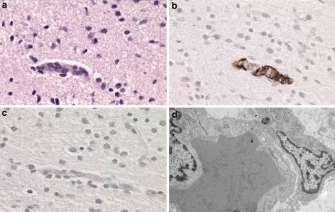
Fig. 1.
Small cerebral vessel (a–c) and small subcutaneous vessel (d) of the foetus with a NOTCH3 mutation. Light microscopy shows a normal vessel wall in the H&E stain (a). Anti-alpha SMA demonstrates the presence of a normal smooth muscle layer (b) and anti-NOTCH3 staining is negative (c). Electron microscopy (×10,000) shows normal structured layers of the vessel, including the endothelial cells and pericyte (right) and some basement membrane material in between, without the presence of GOM or signs of degeneration (d)

