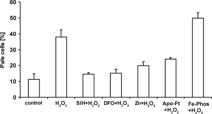
Fig. 3.
Labilization of lysosomes by hydrogen peroxide and its modulation by autophagocytosed metallothioneins and endocytosed apo-ferritin, desferrioxamine and a Fe phosphate complex. J774 cells were exposed to hydrogen peroxide (initially 100 μM in PBS) for 30 min (H2O2), to PBS only (Control), or to H2O2 in combination with 100 μM of the lipophilic iron-chelator SIH—and then returned to standard culture conditions. Some cells had previously been exposed to the hydrophilic iron chelator desferrioxamine (1 mM) for 3 h, to 1 μM apo-ferritin (Apo-Ft) for 4 h, or a Ferric-phosphate complex (30 μM) for 4 h, while other cells had been exposed to ZnSO4 for 12 h to induce the upregulation of metallothioneins and ensuing autophagy of this protein. Note that apo-ferritin, desferrioxamine, SIH, and metallothioneins stabilize lysosomes, while the Fe–phosphate complex has the opposite effect. The figure is compiled from a number of previously published ones

