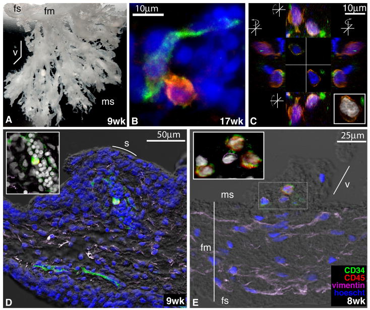
Figure 4.
Localization and morphology of CD34+CD45+ cells within the human placenta. (A) Chorionic villi biopsied from a 9-week human placenta embedded in gelatin to demonstrate the macroscopic anatomy of these structures (2x magnification). fs, fetal side; fm, fetal membranes; v, villus; ms, maternal side. (B) CD34+CD45+ cell within the villous mesenchyme in close contact with a CD34+CD45− _endothelial cell. Tissue sections of the chorionic villi at the gestational ages indicated were stained with anti-CD34 (green) and anti-CD45 (red) mAbs, along with TOTO-3 iodide (blue). Confocal images were obtained by sequential scanning at 63x magnification. (C) Orthogonal views of two CD34+CD45+ cells, stained as in B and from the same 17-week placenta, demonstrating their close associations with CD34+CD45− cells. The Z projection (inset) shows the large size and complex morphology of the putative progenitors’ nuclei. (D) Longitudinal section of a villus showing a single CD34+CD45+ cell surrounded by a cluster of CD34+ CD45− cells. s, syncytium. The tissue section was stained with anti-CD34 (green), anti-CD45 (red), and anti-vimentin (purple) mAbs, Hoescht (blue), and visualized with differential interference contrast (DIC, gray scale). (E) Section of a young villus near the fetal membranes with CD34+CD45+ cells in the center (stained as in D). The Z projection (inset) demonstrates a cluster of CD34+CD45+ cells that appeared to be tethered to their vimentin-positive neighbors.

