Figure 5.
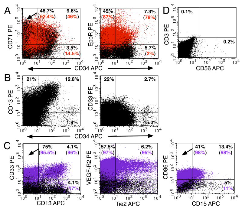
Erythroid and myeloid progenitors together with NK cells were present in the placenta throughout gestation. (A) A freshly isolated light-density cell suspension from a 21-week placenta was stained for erythroid markers and analyzed (see Figure 1 legend). Red indicates cells stained with an anti-CD235a-FITC (glycophorin A) mAb. (B) Analysis of a 12-week light-density placental cell suspension using mAbs that recognized myeloid progenitors in combination with an anti-CD34 mAb. (C) Four-color analysis of antigen expression on mature myeloid CD14+ cells (purple) (same cells as in B). (D) Analysis of CD3 and CD56 expression on 22-week placental cells. In all experiments, 1–2 × 10⁵ PI− cells were analyzed. Results are representative of 5 experiments.

