Fig. 5.

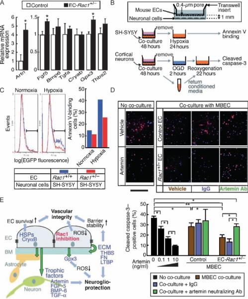
Rac1+/− ECs show enhanced neurotrophic activity through paracrine mechanisms. (A) mRNA expression of the potential neurotrophic factors was assessed in the EC-Rac1+/− and control mouse whole brains by qRT-PCR (n = 7 mouse brains). (B) Schematic of EC-neuron co-culture. (C) Co-culture with Rac1+/− versus Rac1+/+ ECs mitigates hypoxia-induced apoptotic death of SH-SY5Y cells, as assessed by Annexin V-EGFP labeling and flow cytometry. (D) Cortical neurons isolated from fetal mouse brains (∼E16) were co-cultured with EC-Rac1+/− or control MBECs in the presence of artemin-neutralizing antibody (Ab), control IgG, or vehicle in the lower chamber. Control neurons were treated with blank inserts and recombinant artemin proteins. Upper panel, after OGD and reoxygenation, the neurons were double-labeled with DAPI (blue) and cleaved caspase 3 (red). Scale bar, 1 mm. Lower panel, quantification of the cleaved caspase 3–positive fractions. (E) A proposed model of neuroprotection through targeting endothelial Rac1. Values are means ± SEM (*P < 0.05, **P < 0.01; ANOVA).
