
Figure 4.
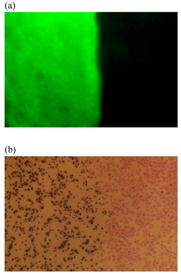
Anti-DIG IgG immobilization was spatially controlled by a low melting point wax masking technique. (a) Anti-sheep IgG antibody conjugated FITC was used to label the surface antibody. Only the exposed area without wax protection illustrated green fluorescent expression. (b) HGF cells were cultured on chitosan surfaces for 2 days. The transduced cells turned blue after X-gal staining. Cells grew to confluence on the material surfaces; however, cell transduction was restricted to the non-masked area. These findings are consistent with the results of the fluorescent labeling.