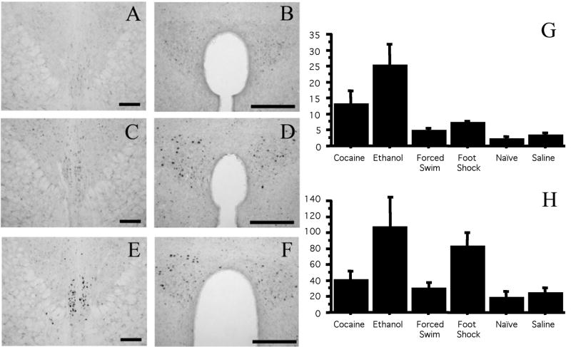
Figure 5.
Fos-ir after administration in pIIIu and the paraventricular nucleus of hypothalamus in naïve mice or mice injected with saline, 20 mg/kg cocaine, 2.5 g/kg ethanol, or exposed to footshock or swim stress (N=6 per group). DAB staining of the pIIIu revealed lack of Fos-ir in the naïve, saline, footshock and swim stress condition, but a significant increase in Fos-ir after the cocaine and ethanol treatment. Representative staining in the pIIIu (A, C, E) or paraventricular nucleus of hypothalamus (B, D, F) in the naïve (A, B), footshock (C, D) and cocaine (E, F) groups is shown on the top. Bar=100 μm. Quantification of induction across conditions in pIIIu is summarized in (G), Quantitation across conditions in the paraventricular nucleus of hypothalamus is summarized in (H).

