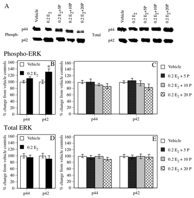
Figure 2.
(A) Representative Western blot images illustrating phosphorylated and total p42 and p44 ERK protein levels in each group. (B) Densitometric analyses indicated that phospho-p42 ERK levels in the dorsal hippocampus were significantly increased 1 hr after a single i.p. injection of 0.2 E2 relative to vehicle controls (* P < 0.05). (C) In contrast, none of the groups treated with 0.2 E2 + progesterone exhibited increased phospho-p42 ERK levels. Phospho-p44 ERK protein levels were not significantly elevated by 0.2 E2 alone or any combination of 0.2 E2 + progesterone. (D and E) No treatment affected total p42 or p44 ERK protein immunoreactivity. Each bar in B–E represents the mean percent change from vehicle controls (± S.E.M.).

