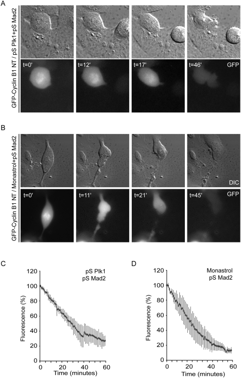
Figure 1. APC/C-Cdc20 Activity in Plk1-depleted U2OS cells.
A–B U2OS cells were transiently transfected with 1 µg of GFP-Cyclin B1-NT, 10 µg of pS-Plk1 and 10 µg of pS-Mad2 as indicated. 18 h after transfection, cells were incubated for 24 h in thymidine. 10 h after washing away thymidine, cells were transferred to the heated stage of a time-lapse microscope. At indicated time points, DIC images and fluorescent light were analyzed. Directly after washing away thymidine, monastrol was added to culture medium, where indicated. C, D Fluorescence levels were quantified using Metamorph software (n = 5 for each condition). Quantified images were plotted from the time of mitotic entry as define by nuclear envelope breakdown (t = 0) and the standard error of the mean of 5 experiments is indicated.

