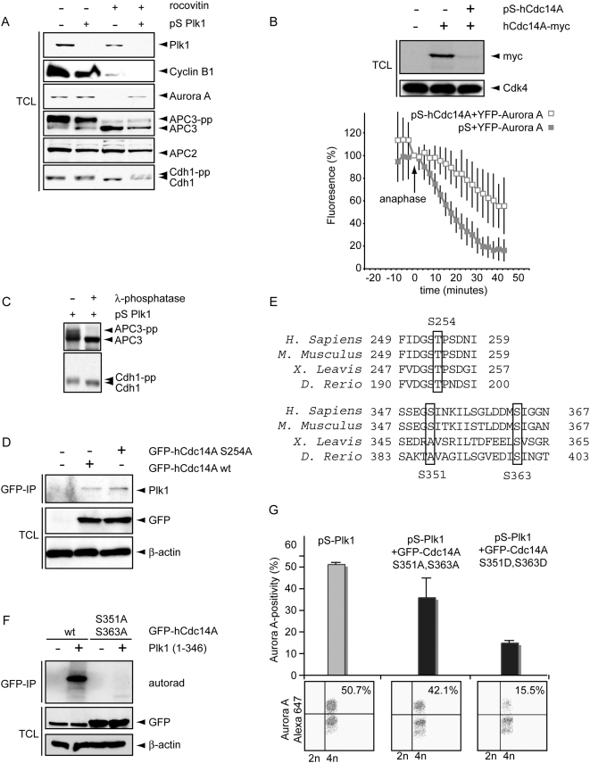
Figure 5. Plk1 controls Cdc14A to regulate the APC/C-Cdh1.
A. U2OS cells were treated with pS or pS-Plk1 as in Figure 4B. Cell lysates were obtained after 2 h of Roscovitine treatment. Total cell lysates were blotted for Plk1, Cyclin B, Aurora A, APC3, APC2 and Cdh1. B. U2OS cell were transfected with hCdc14A-myc in combination with pS-hCdc14A. Cell lysates were immunoblotted for Cdk4 and myc. In parallel, U2OS cells were transfected with Aurora A-YFP A in combination with pS or pS-hCdc14A. After release from a thymidine block cells were analyzed using live microscopy. Aurora A levels were quantified and mean and SEM of 5 movies were synchronized at anaphase onset. After anaphase onset, 50% degradation of Aurora A was reached on average after 41 minutes for pS-Cdc14A transfected cells versus 17 minutes in control cells. C. U2OS cells were transfected with pS-Plk1and mitotic cells were collected by mitotic shake-off 60 h after transfection, and subsequently lysed. Extracts were incubated in the presence or absence of lambda-phosphatase. Western blots were probed with anti-APC3 or anti-Cdh1. D. U2OS cells were transfected with wt-GFP-hCdc14a or GFP-hCdc14A-S254A. After 48 h, GFP-immunoprecipitations were performed and total cell lysates and GFP-IP's were immunoblotted for GFP, Plk1 and β-Actin. E. The conservation in the regions comprising Ser254, Ser351 and Ser363 of Cdc14A is indicated. F. U2OS cells were transfected with wt-GFP-hCdc14a or GFP-hCdc14A S351, 363A. After 48 h, GFP-immunoprecipitations were performed. GFP-immunoprecipitations were left untreated or incubated with recombinant Plk1. Kinase reactions were visualized by autorad. Total cell lysates were immunoblotted for GFP and β-Actin (lower panels). G. U2OS cells were transfected with pS-Plk1 in combination with GFP-hCdc14A S351,363A or GFP-hCdc14A S351,363D. 36 h after transfection, nocodazole was added to cell cultures. After 16 h, mitotic cells were collected by shake-off. Mitotic cells were subsequently replated in presence of Roscovitine for 4 h, and subsequently cells were fixed in ethanol and stained with anti-Aurora A-Alexa-647. Representative Aurora A-plots of GFP-positive cells are shown (lower panel) and the mean and SEM of 3 experiments are plotted (upper panel).

